A radioisotope emitting positrons is given to a patient intravenously; PET allows to follow the journey of the radioisotope in the body and in the organ under examination. PET is the most sensitive and specific technique to visualize the journeys and the interactions of molecules during the physiological activity. The good selectivity of the tracer allows to use very small doses of radioactivity. PET is used to study the functional anatomy of various neurophysiological activities of the brain. Neuropsychiatry and neurology use this method to improve the knowledge of the fisiopathology of the brain. PET is also used for the study of brain tumours and to detect mechanisms of antitumoral therapies. It may be extremely powerful for the development of new drugs.
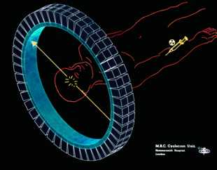
1) Scheme of PET: a nuclide in a drug emits a positron, which slows down, interacts with an electron yielding two photons of fixed energies, in opposite directions The two photons are detected contemporarily, thus allowing the localization of the radionuclide in the tissue under investigation.
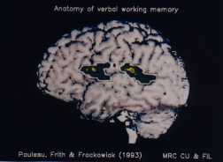
2) Application of PET to neurofisiology: the activated areas (in colour), detected by PET, are the brain regions in which memorization procedures are realized, as when we try to remember a telephone number.